Fragment analysis
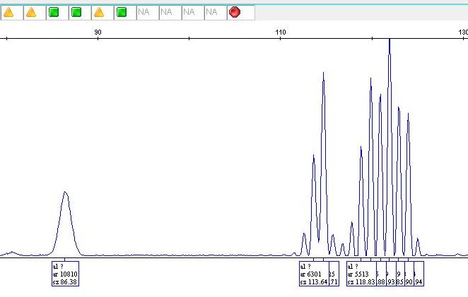
After certain DNA fragments have been amplified, fragment analysis makes it possible to visualise their length by means of capillary electrophoresis. This technique is used when diagnosing lymphoproliferative disorders (clonality analyses) and identifying microsatellite instability.
Microsatellite instability (MSI)
Microsatellites are short, non-coding DNA sequences, ranging in length from 2 to 6 base pairs, that are often repeated within an organism’s genome. With microsatellite instability (MSI), defective DNA repair proteins (hMLH1, hMSH2, hMSH6, hPMS1, hPMS2) cause changes in length within these microsatellites. If microsatellite instability is identified in the tumour under examination, it can be assumed that the patient has a genetic defect in their DNA repair system. This genetic defect may be present only in the tumour (sporadic) or, in rare cases, could be inherited (mutation in the germline). In such cases, genetic counselling and extended family screening are advisable.
Patients with colorectal cancer are often tested for microsatellite instability to rule out hereditary non-polyposis colorectal cancer (Lynch syndrome), amongst other things.
Clonality analyses
Clonality analysis is used to support the diagnosis of B- or T-cell lymphomas. This method is based on the unique gene rearrangement of B-cell receptors (Ig) and T-cell receptors (TCR) in B- and T-lymphocytes. The length of the modification varies between rearranged gene combinations of different lymphocytes in reactive processes (polyclonal pattern), but not in neoplastic lymphocytes which represent clonal derivatives of a single transformed cell (monoclonal pattern).
Your contact person
PD DrDavide Soldini, Head Pathology
Head Pathology, Head molecular pathology
Specialist in Pathology and Molecular Pathology FMH
DrSimone Brandt
Deputy Lead Molecular Pathology
Specialist in Pathology and Molecular Pathology FMH


Our Molecular Pathology is an SIWF certified continuing education center.

